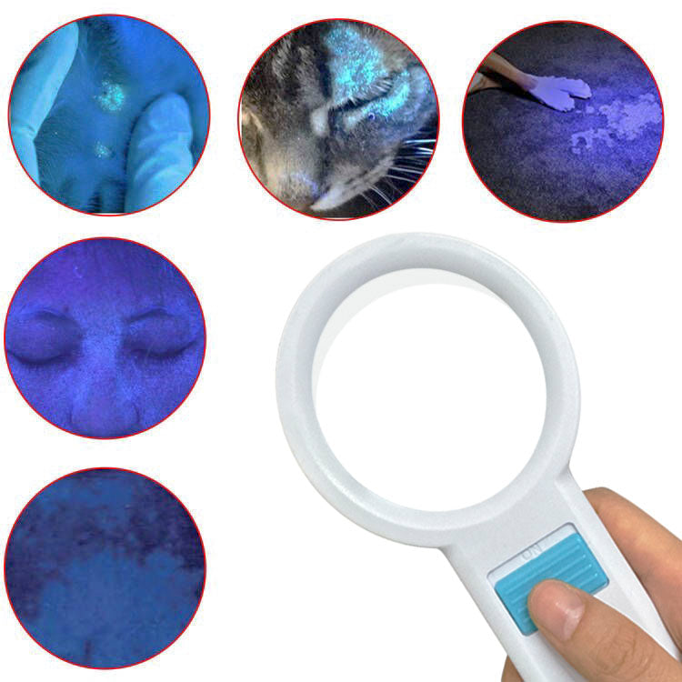
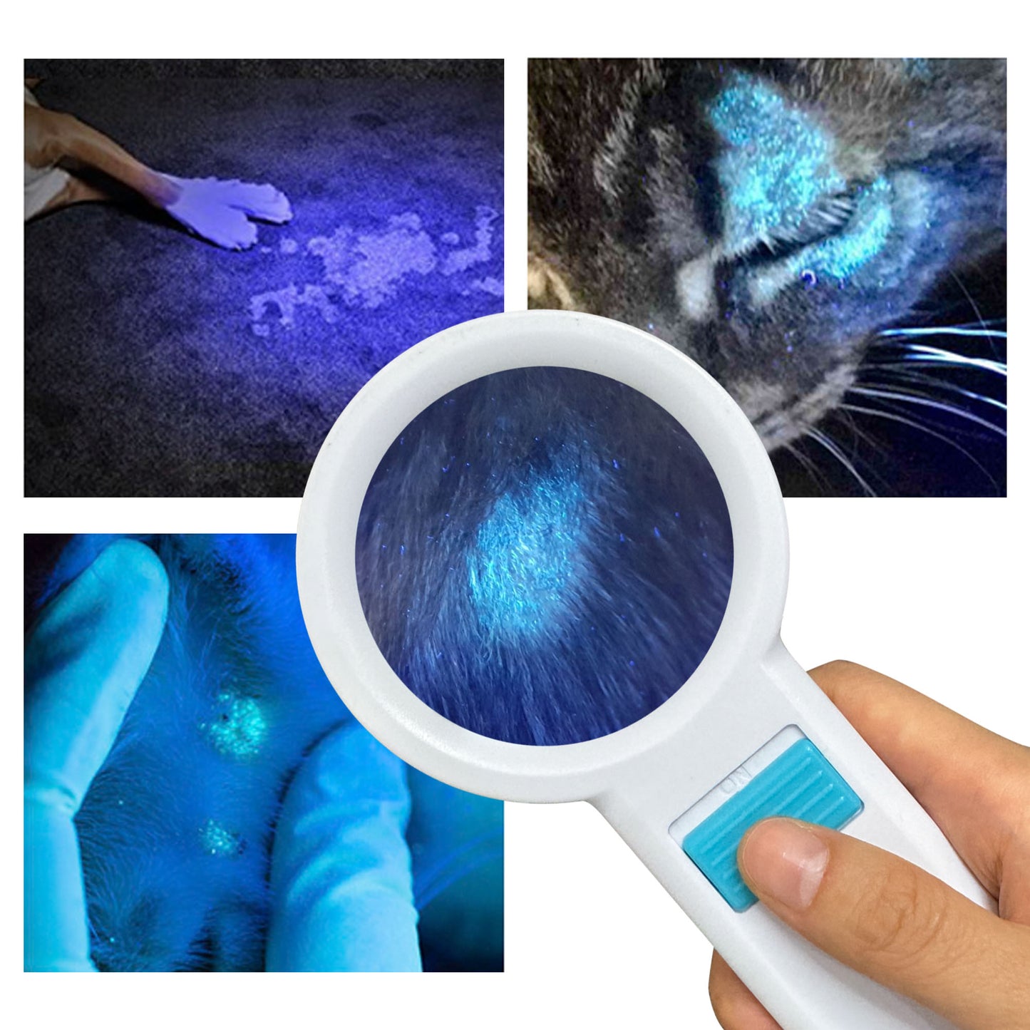
What skin conditions can a Woods lamp help diagnose?
Featured collection
-
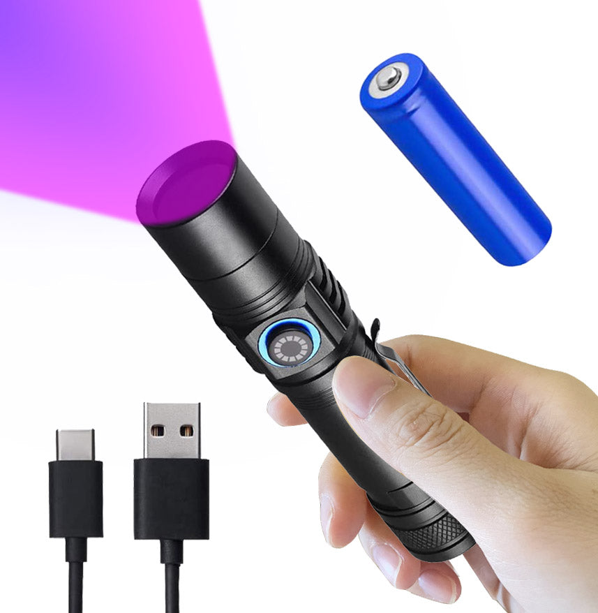
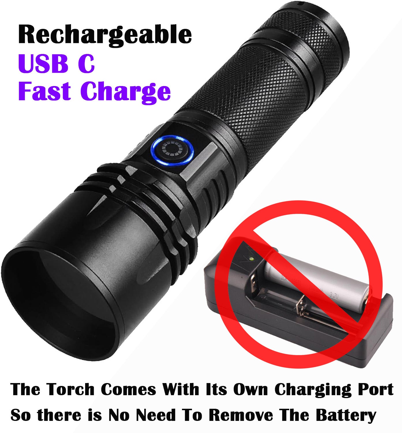
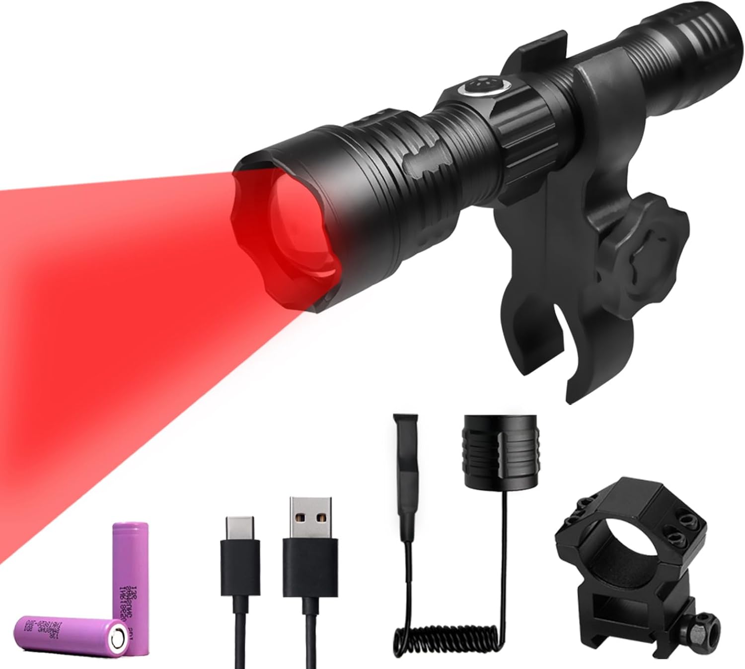
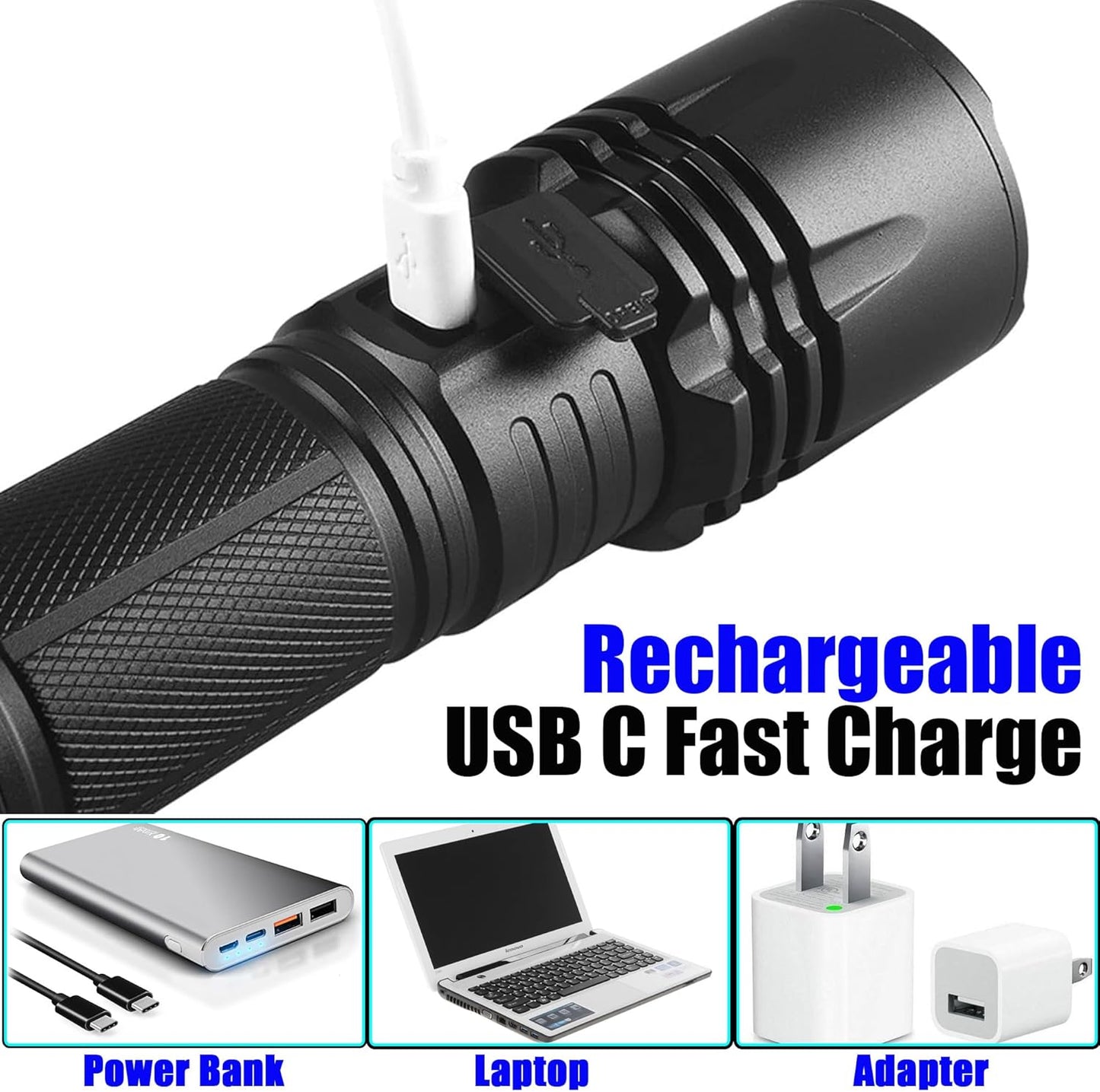
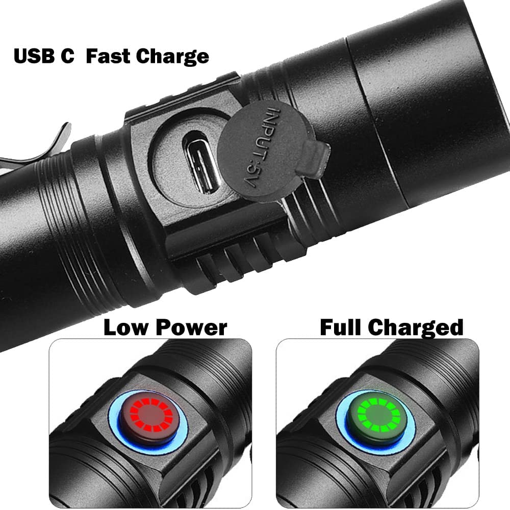
Uv Blood Tracking Flashlight for Hunting Deer Finder Blood Trailing Light Rechargeable Blood Tracker Light
Precio habitual A partir de $29.99Precio habitualPrecio unitario por$79.99Precio de oferta A partir de $29.99Oferta -
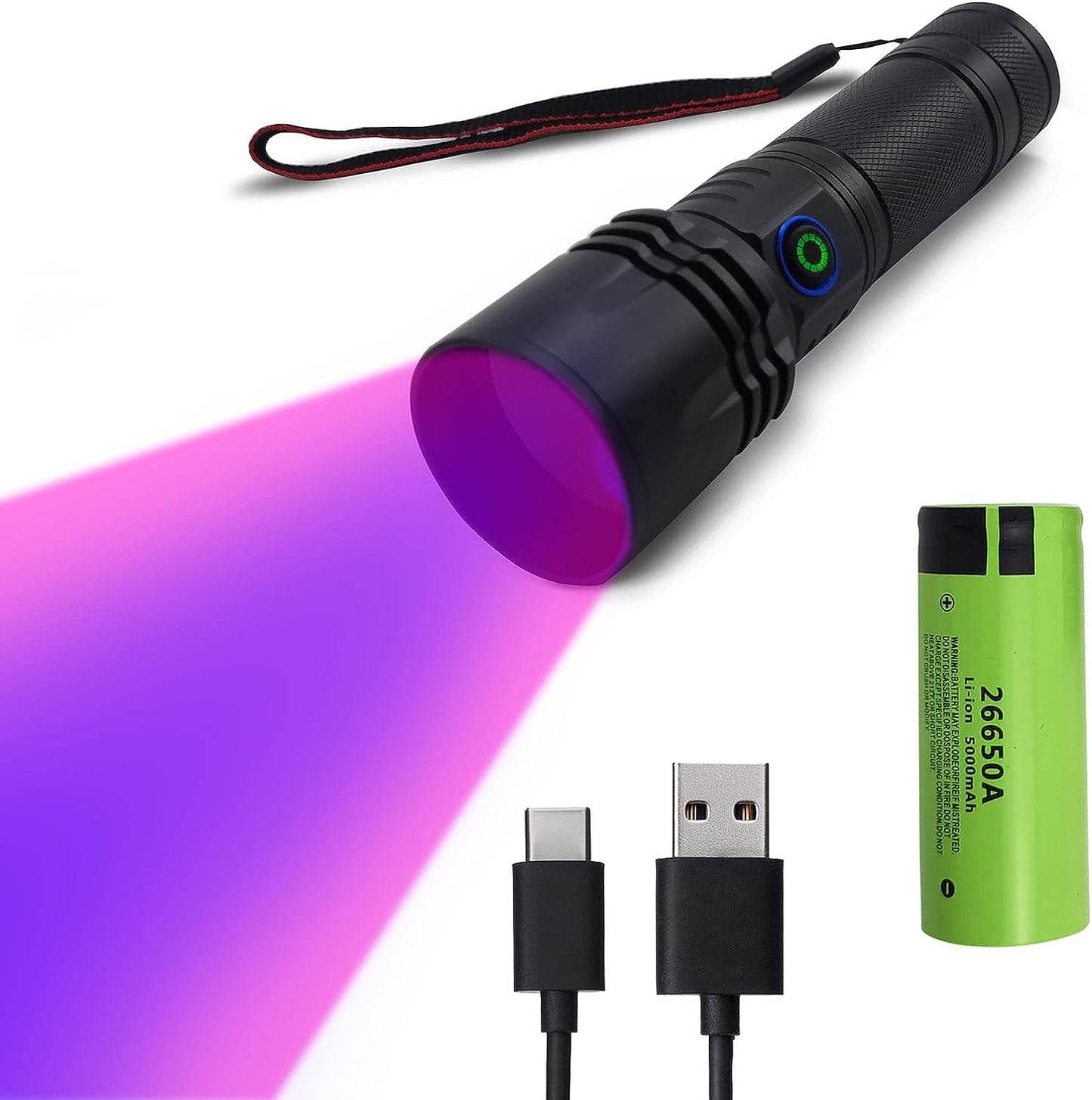
Rechargeable Blood Tracking Light for Night Hunting 2000 Lumens Blood Trail Tracking Flashlight Gifts for Hunter (Blood Finder Light)
Precio habitual $39.99Precio habitualPrecio unitario por$69.99Precio de oferta $39.99Oferta -
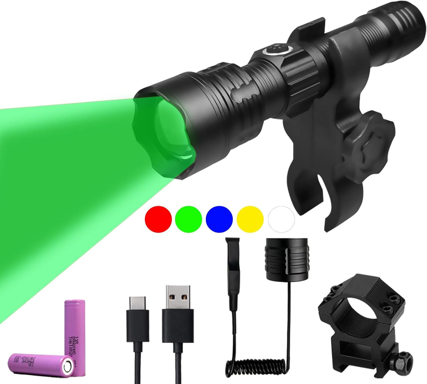
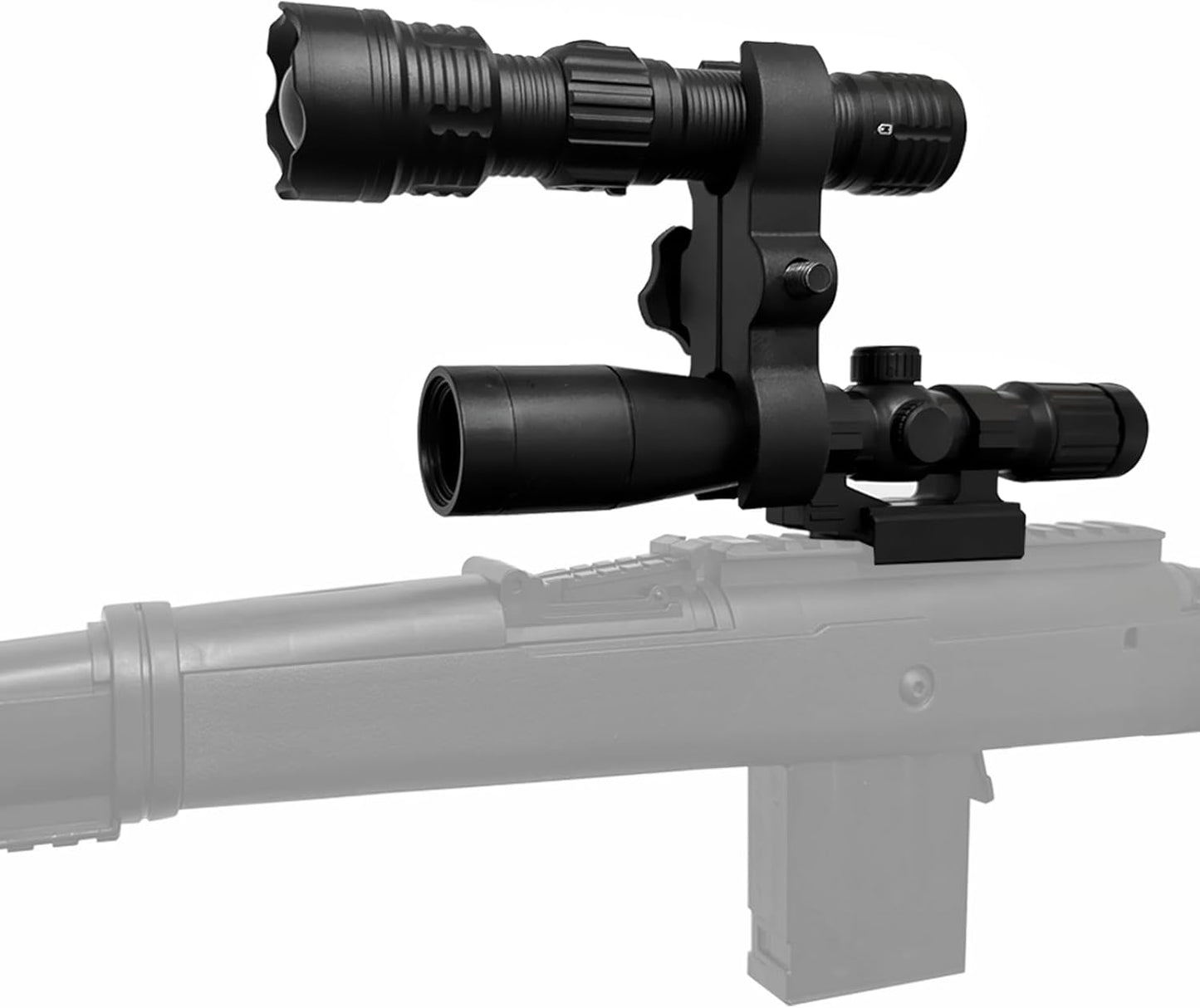
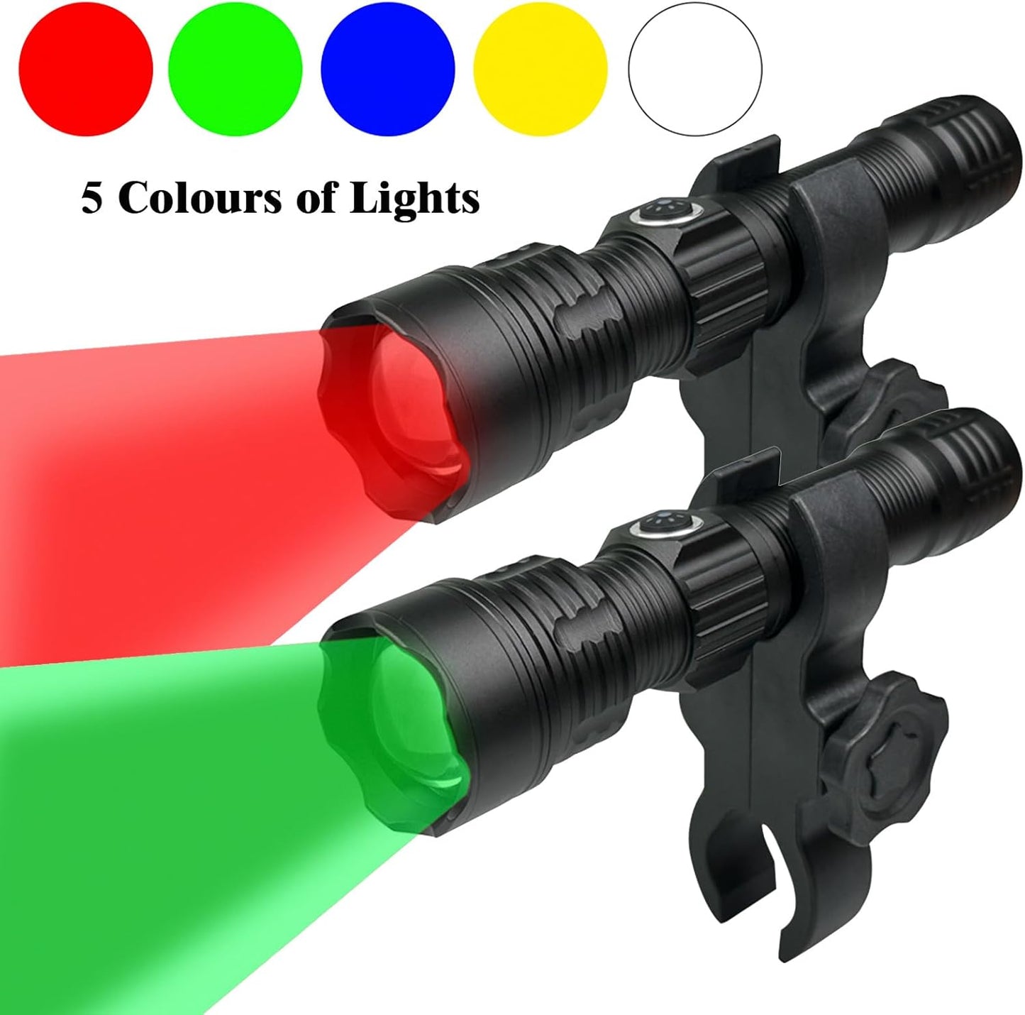
Green Light for Hunting Hog Green Flashlight,Red,Blue,White 4 in 1 Light for Coyote,Hog,Coon,Predator,Varmint,Sniper,Scope,Hunting Lights (Hog Green Light)
Precio habitual $79.99Precio habitualPrecio unitario por$199.99Precio de oferta $79.99Oferta -
Coyote Hunting Light,Red Light for Hunting,Red,Green,Blue,White 4 in 1 Light for Coyote,Hog,Coon,Predator,Sniper,Scope,Hunting Lights
Precio habitual $79.99Precio habitualPrecio unitario por$199.99Precio de oferta $79.99Oferta -
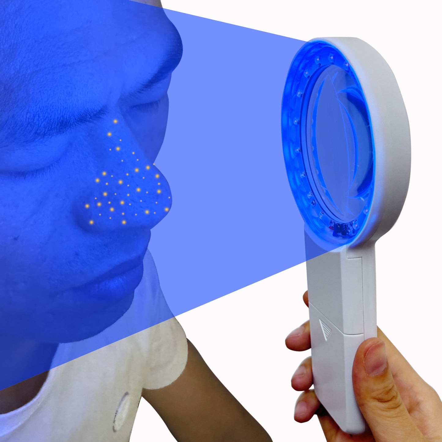
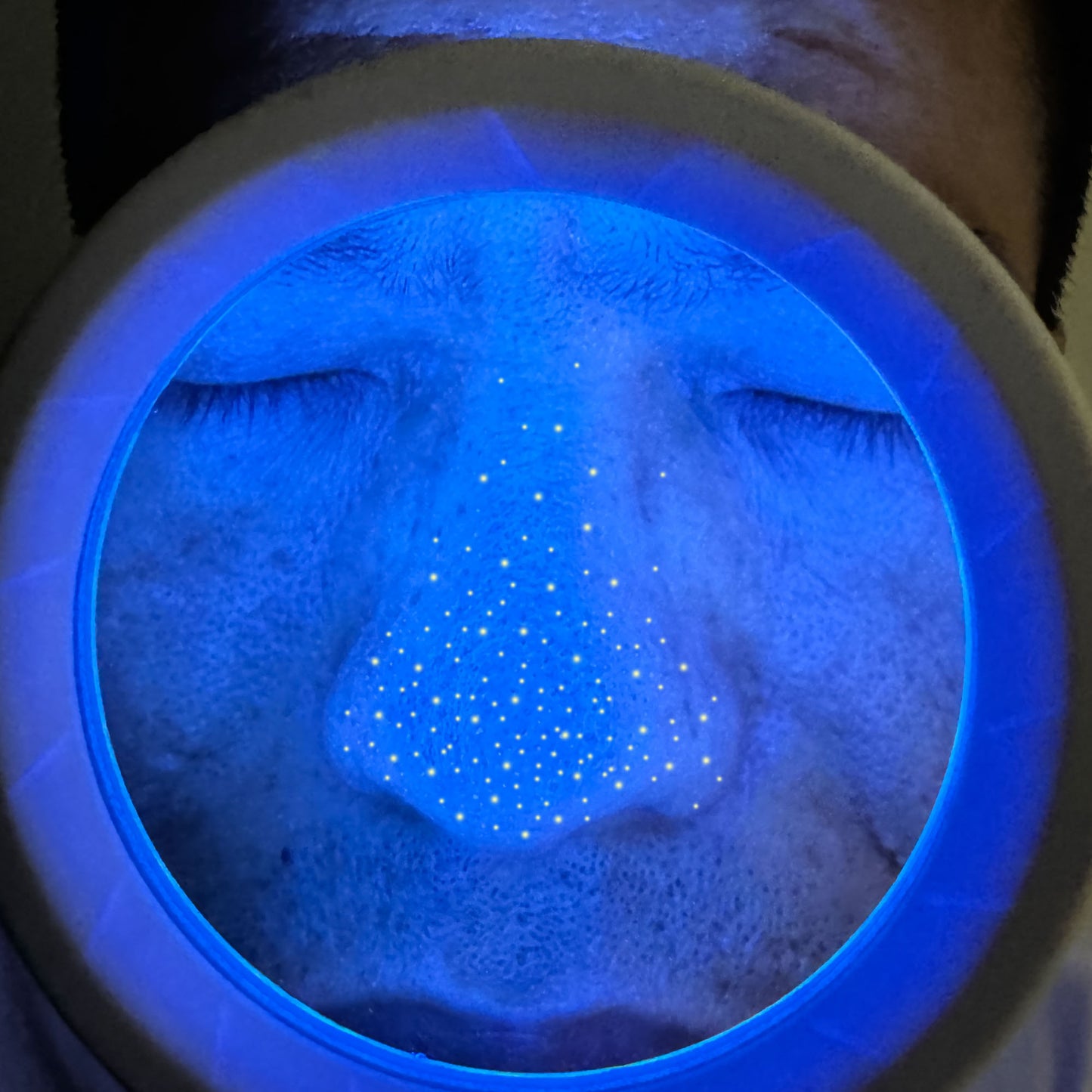
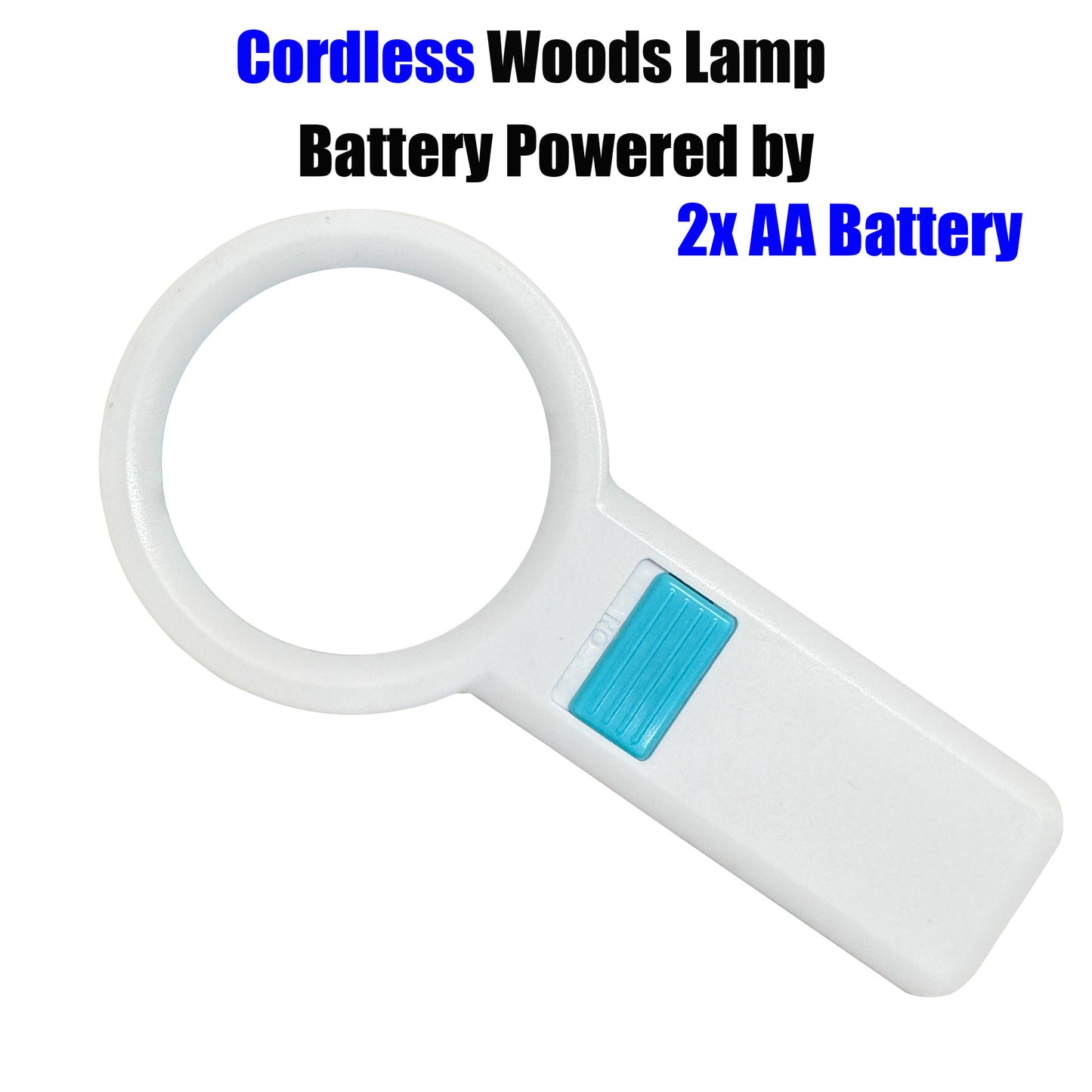
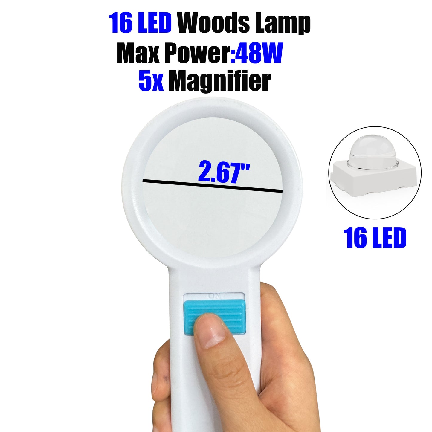
Cordless Wood's Lamp Ringworm Detection Light-Skin Testing-Esthetician-Veterinaria-5x Magnifying Wood Lamp Black Light-16 LED-Battery Powered Polarized Skin Dermatology Dermascope Light
Precio habitual $49.99Precio habitualPrecio unitario por$99.99Precio de oferta $49.99Oferta -
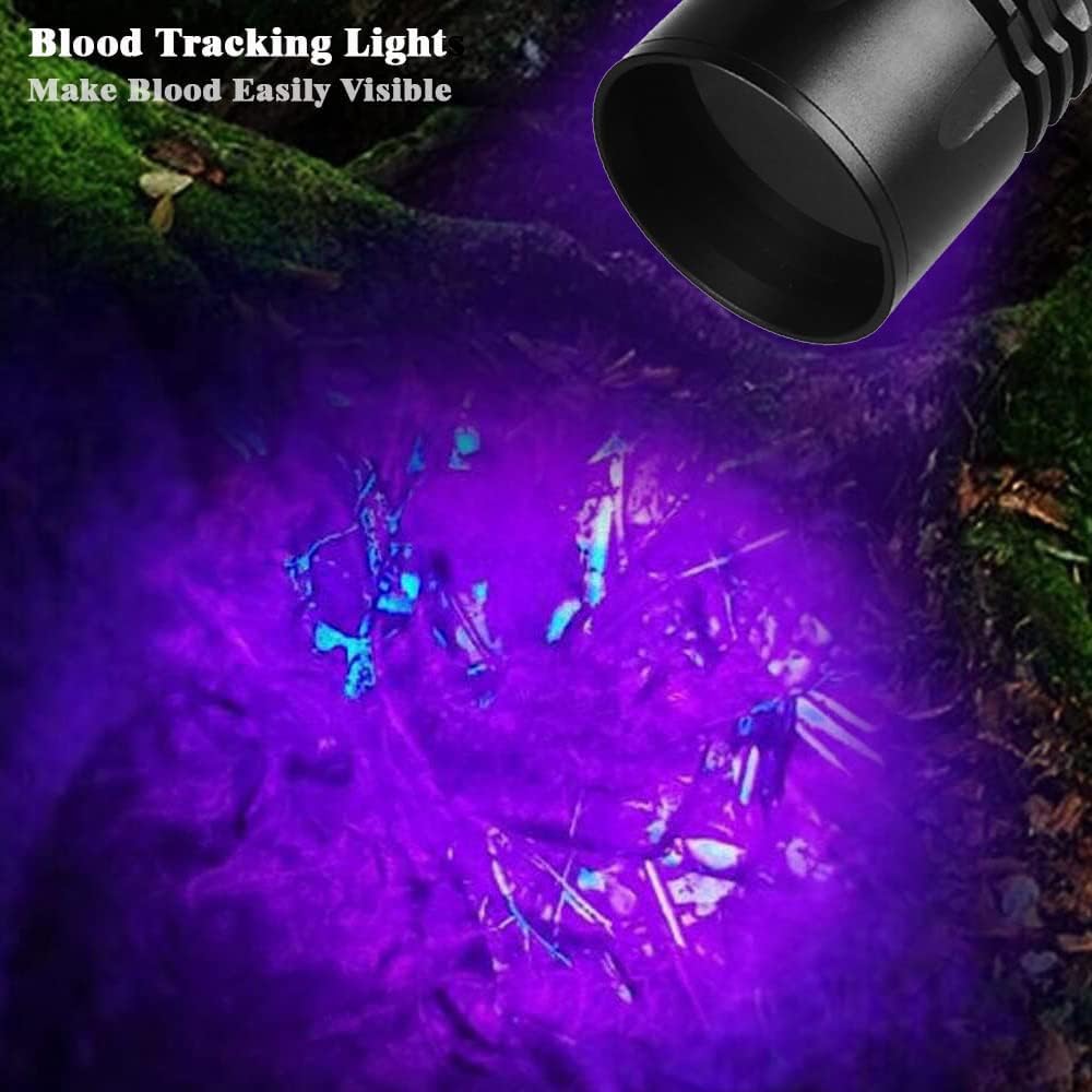
Best Rechargeable Blood Tracking Flashlight for Hunting Deer Blood Trail Finder At Night Perfect for Wounded Game Tracking-Hunters Gifts
Precio habitual $39.99Precio habitualPrecio unitario por$69.99Precio de oferta $39.99Oferta -
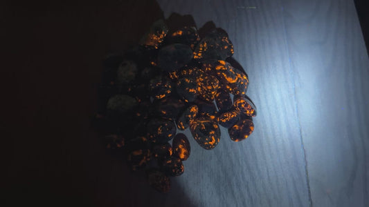
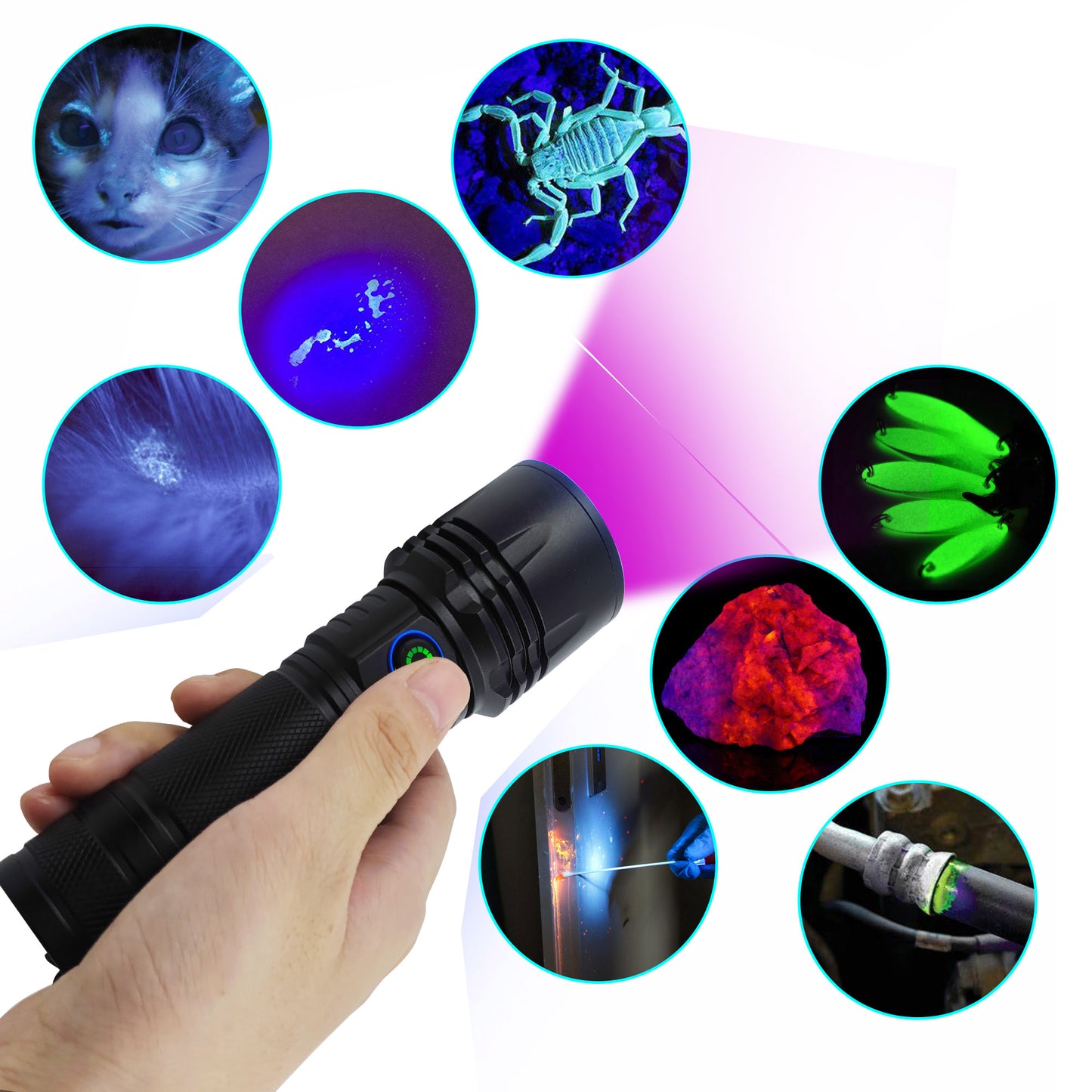
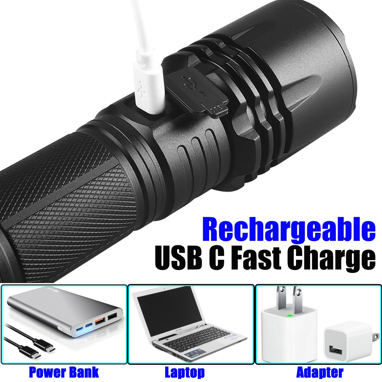
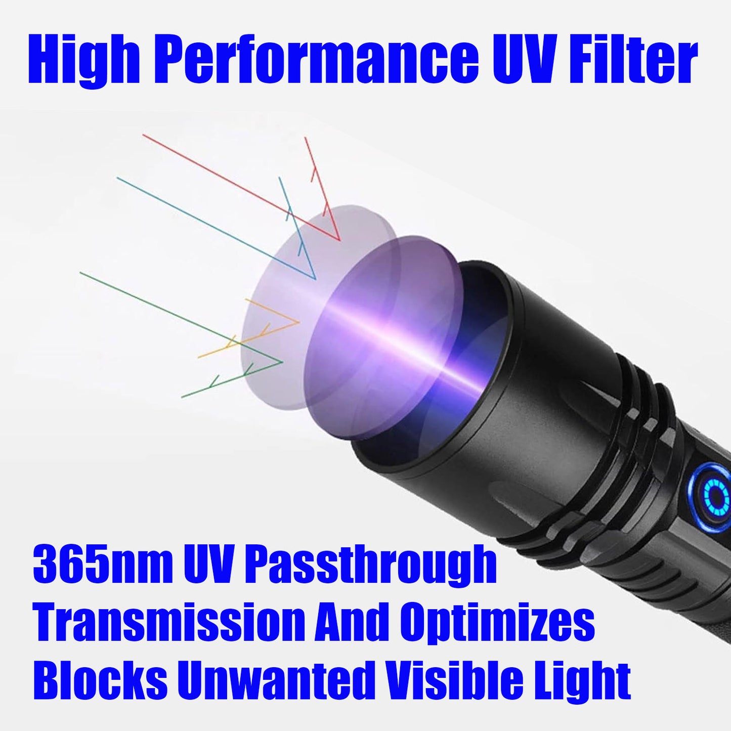
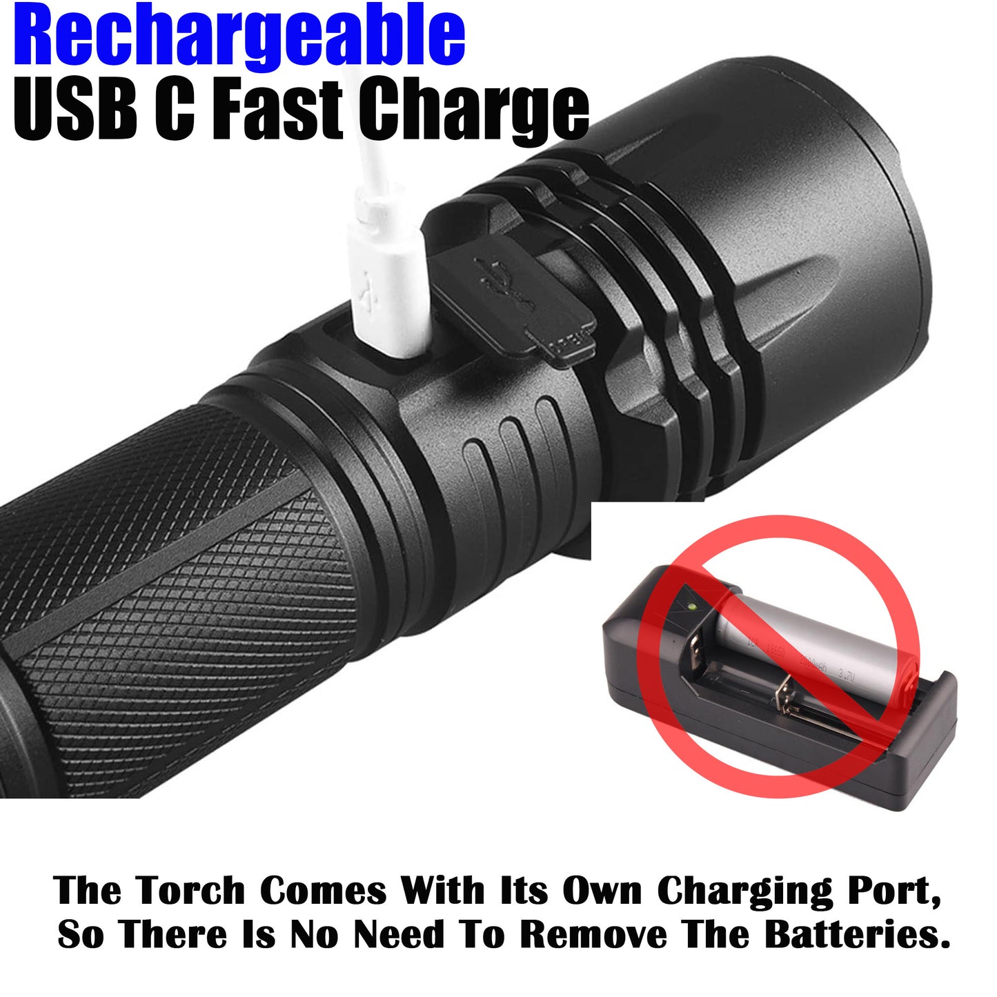
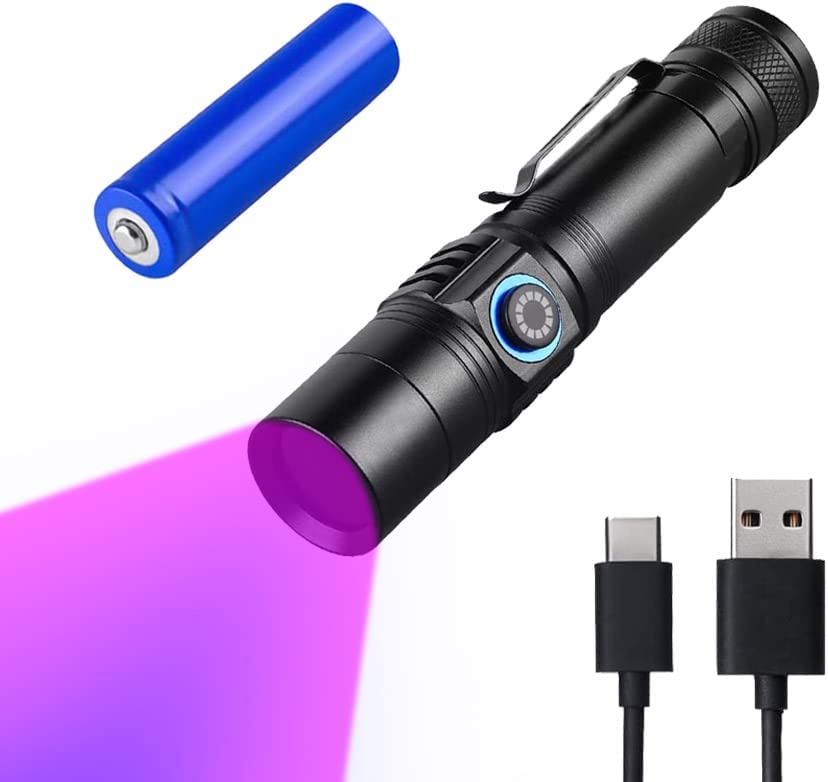
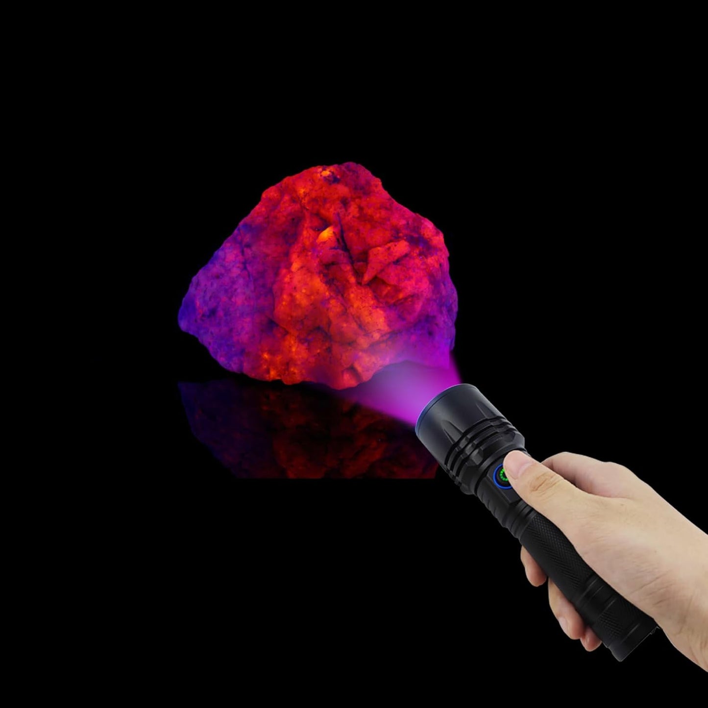
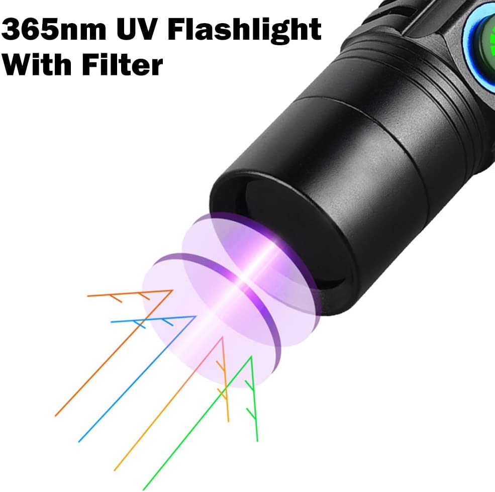
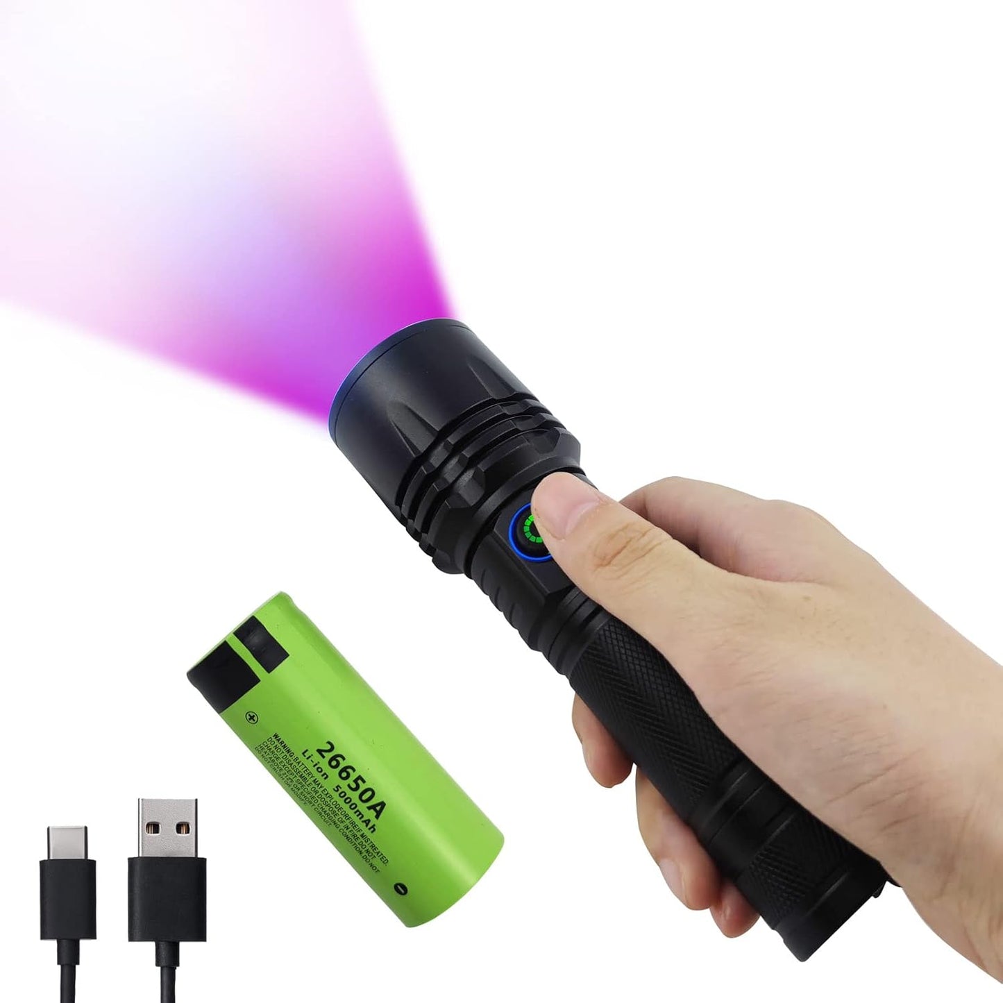
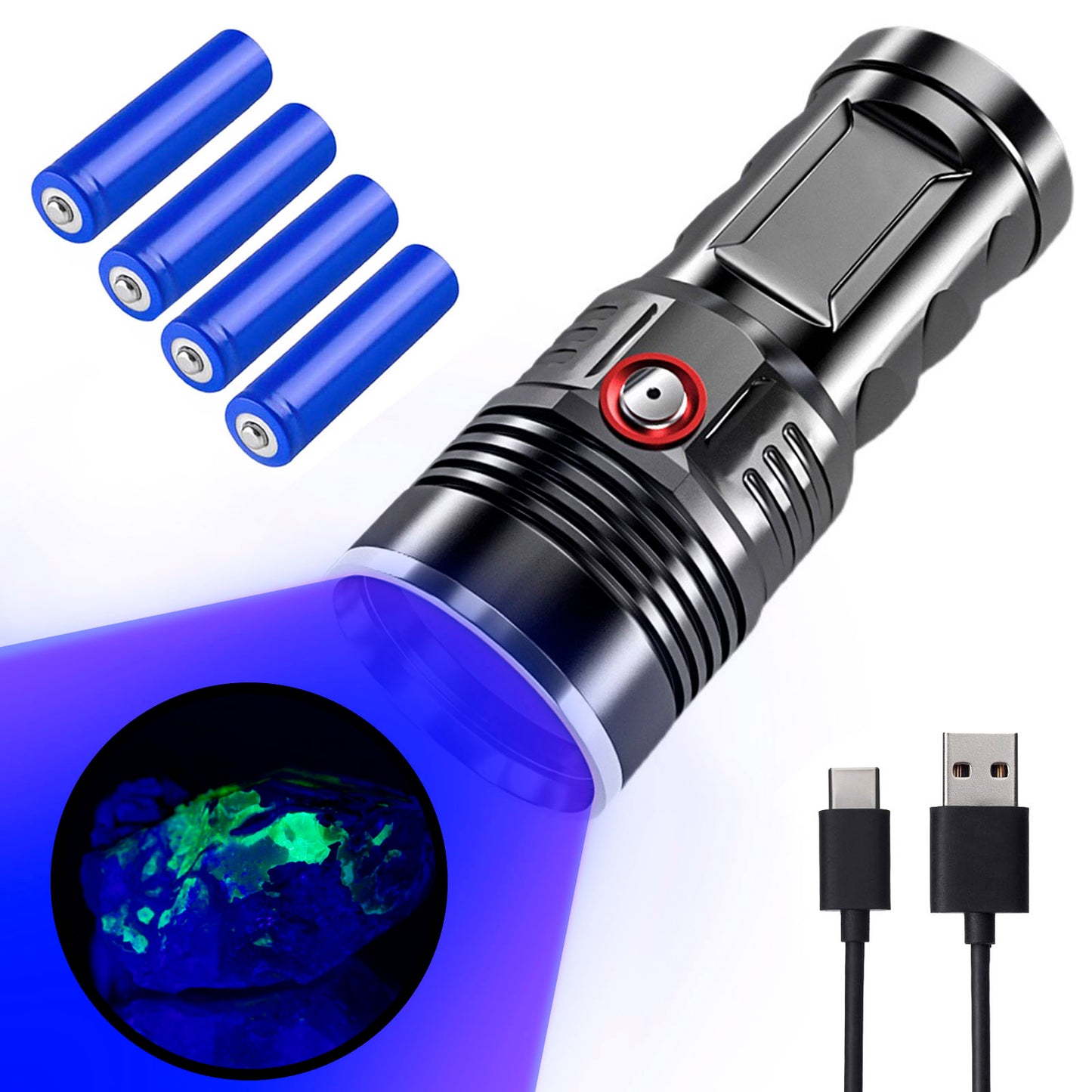
365nm UV Flashlight for Rock Hunting & Mineral Detection - Professional Gemstone Detector Tool with High Power Short/Long Wave, Portable UV Light for Crystals, Agates, Uranium Glass, Jade Appraisal
Precio habitual A partir de $29.99Precio habitualPrecio unitario por$79.99Precio de oferta A partir de $29.99Oferta